COVID-19 swept across Georgia in early spring, a deadly force flattening life’s landscape. All of Emory Healthcare, including Emory Radiology, swiftly implemented protocols to save lives while preventing infection spread. Life-saving work happened behind closed doors by team members whose dedication shone through their eyes, the only part of them visible beneath layers of personal protective equipment.
The following describes just some of the ways every member of the department united in sacrifice and commitment, rising together with hope for a better tomorrow while fighting to save lives and protect health today.
The following describes just some of the ways every member of the department united in sacrifice and commitment, rising together with hope for a better tomorrow while fighting to save lives and protect health today.
REDEFINING THE WORKPLACE |
RECONFIGURING OPERATIONS |
Immediately transitioning to remote working and learning not only limited virus transmission, it also freed space to be reconfigured as needed for COVID care.
|
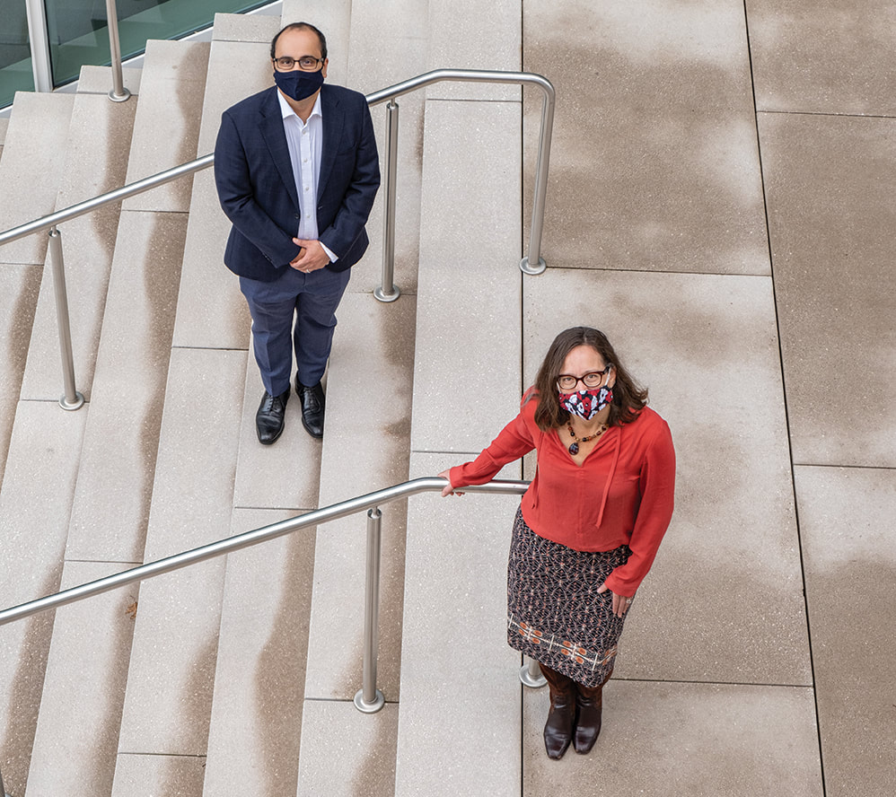
Clinical operations leaders including Nursing Director of Radiology & Imaging Services Pamela Stanley, MSN, RN, PCCN-K, and Dr. Marta Heilbrun worked with Emory Healthcare Infection Prevention and Control to reconfigure clinical operations
|
TRAINING IN THE ONLINE HOT SEAT |
SAFELY RESUMING CLINICAL ROTATIONS |
|
Ryan Peterson, MD, assistant professor and associate program director of the Diagnostic Radiology Residency, pivoted to Zoom for case conferences. The resident in the “hot-seat” would prepare the case and then answer questions and provide recommendations for next steps. High-yield cases helped first year residents prepare for call; more complex cases challenged upper-level residents. Brent Weinberg, MD, PhD, assistant professor, answered questions and added teaching points using the “chat” function. Chief Resident Patricia Balthazar, MD, PhD, promoted case sessions on social media and managed participant access to prevent security breaches. The sessions proved so popular, faculty and trainees from Indiana University, University of Florida, Georgetown University, Medical College of Wisconsin, University of South Florida, and AdventHealth in Orlando swelled attendance from 50 to 120 learners, creating a rich environment for collaborative learning.
|
The undergraduate medical imaging program resumed clinical training in phases starting in June. Every student and faculty member received PPE kits and access to on-demand COVID testing. Students also were protected from encountering COVID+ and potentially COVID+ patients. Seniors returned June 1 to complete clinical rotations and other requirements with Emory covering tuition so they could graduate in July. Clinical rotations for all other BMSc students resumed on June 29; simulation labs reopened August 1 with fewer students per section and more sections. Didactic classes continued online like most of Emory’s undergraduate classes in the fall.
“The pandemic pushed us to make tough choices but we remained committed to ensuring our students could fulfill their academic requirements without jeopardizing either their safety or our faculty’s safety,” says Program Director Theodore Brzinski, Jr., MS, RT(R). |
FUSION ARCHITECTURE TRAINED FOR COVID
Imon Banerjee, PhD, Judy W. Gichoya, MD, MS, and Hari Trivedi, MD, assistant professors in Emory Radiology’s Health Innovations and Translational Informatics Lab, are collaborating with colleagues at Stanford University on a model that fuses patient clinical data, including risk factors, with imaging studies to predict COVID diagnosis and disease progression. By training the model using clinical data from highly diverse patient populations both with and without COVID, the investigators aim to create a powerful tool to expedite diagnosis and treatment planning to maximize patient outcomes. The project leverages the expertise of Drs. Banerjee and Gichoya in fusion architecture—incorporating data from many sources—for deep machine learning to predict clinical outcomes.

PREDICTING COVID PULMONARY SEVERITY |
UNDERSTANDING IMPACT ON PATIENT VOLUME |
|
Carlo De Cecco, MD, PhD, associate professor, and Marly van Assen, PhD, post-doctoral fellow, are collaborating with Ali Adibi, PhD, director of the Center for Advanced Processing-tools for Electromagnetic/acoustics Xtals at Georgia Tech, on the COVID-19 PREDICTION Study. Submitted as an R21 NIH grant, the project aims to build a predictive model that can distinguish COVID-19 pneumonia from other lung pathologies using chest imaging and clinical data. Machine learning also will monitor disease progression over time, ideally finding imaging and clinical parameters that can identify patients likely to develop severe cases of lung-involved COVID-19. They hope this tool for COVID-19 early detection and prognostication will aid clinical decision-making to improve patient outcomes and optimize resource allocation. Initial results are described in a paper titled “Toward Understanding COVID-19 Pneumonia: A Deep-learning-based Approach for Severity Analysis and Monitoring the Disease,” submitted to the peer-reviewed journal Nature Biomedical Engineering.
|
Dr. Jamlik-Omari Johnson and colleagues are examining the impact of COVID-19 infection fears on patient care-seeking in emergency departments. The results are guiding the team in developing strategies to address delays in care as COVID infections continue to surge.
Dr. Richard Duszak, Jr., vice chair for policy and practice, is evaluating the impact of the shutdown on imaging volumes from nine community radiology practices across the US. Diagnostic imaging volumes substantially declined, especially in mammography, suggesting a next phase of research about the impact of delayed medical care on patient health. |